Biology (Morphometrics) and Paleontology Applications for 3-D Data Collection and Analysis
Biology and Paleontology Applications
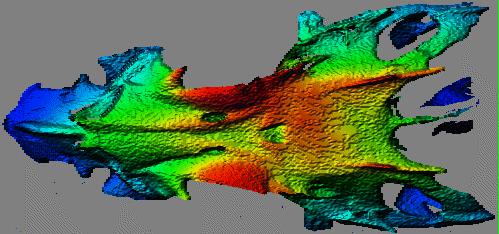
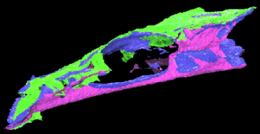
As part of a collaborative effort with the Gulf Coast Research Laboratory in Ocean Springs, Mississippi, studies have determined the feasibility of measuring fish skeletal features using these techniques. This measurement technique has much promise for the related fields of biological systematics, physical anthropology, and morphometrics. Also see the page on integrating data from multiple viewpoints for more icthyological results.
3-D measurements are typically limited to a relatively small collection of inter-landmark distance gathered by hand-held calipers. CT or MRI offer some promise, but they are expensive sensing modalities. Our technique gathers a rich data set in a relatively short time at a low cost. Not only can the traditional inter-landmark distances be measured from a range map, but researchers could now analyze 3-D space curves, or even 3-D surfaces. Consider the load-bearing surface of a joint — the area of that gives some indication of how much load the limb bore, and the 3-D shape of the surface may indicate how the limb was used.
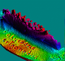
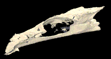
The first image below shows the mandible, or lower jaw, of Pelycodus trigonodus, a small lemur-like primate that inhabited present-day North America and Europe about 50,000,000 years ago. The visible portion here is about 32 mm long; the molars, on the right, are about 2.5 mm tall and 3 mm wide. The target is a cast of a specimen in the collection of the American Museum of Natural History in New York.
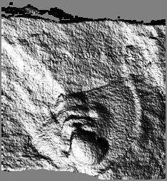
The last image above shows a trilobite fossil from about 530 million years ago. The exposed part of the specimen is approximately 15.5 mm wide by 10.5 mm long, with total relief limited to perhaps 1.5 mm. Click here or on the image for more trilobite images.
There are also some range maps of fossilized insects. Click here to see those images.
