The New Range Sensor
The New Range Sensor
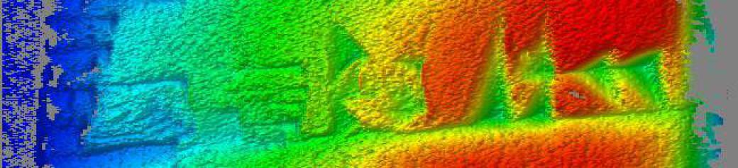
The new range sensor is now in operation at the Gulf Coast Research Laboratory in Ocean Springs, Mississippi. Here it is in action, scanning a sample:

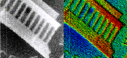
Test Images
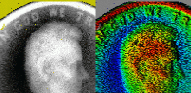
Let's look at the most impressive result first. Here are parts of the front and rear of a penny.
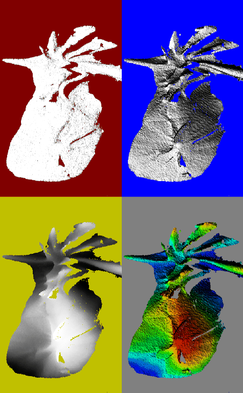
Now for the first three infraorbital bones of Scorpaena plumieri. This is 60 mm long by 20 mm wide by 7 mm deep.
The upper left image is registered intensity.
The upper right image is rendered 3-D shape.
The lower left image is depth-as-intensity.
The lower right image uses rendered 3-D shape for intensity, and color-encodes depth as hue.
Click here or on the image at left for a detailed image.
A small valley can be seen running left-to-right, about 40% of the way from the bottom to the top in the two subimages seen at right above. Below is a more detailed view from the image using rendered 3-D shape for intensity and hue for depth.

It starts near the left edge, and continues at least 60% of the way across the specimen, just above a V-shaped formation. That valley is a structure on the same scale as a suture. I don't think it is a suture, but then again I don't know enough about icthyological anatomy to know if the lacrimal bone is a collection of smaller bones joined by sutures, as the skull is. At any rate, this shows promise that at least on skulls of moderate size, say at least 40 mm long, sutures may be detectable by 3-D shape alone. I was later told:
The lacrimal is composed of osteological centers that evidently fuse. It would appear that the lacrimal in fishes is actually composed of the consolidation of several (as I recall 3) growth centers, only one of which is evidently homologous to the lacrimal that one sees in mammals. To be honest I'm not sure exactly which of the several ridges on the first infraorbital bone (lacrimal) you are looking at but it is remarkable that you even picked up on this.
What you are looking at in the scan is the first three infraorbital bones, the "lacrimal" being the first. Its the one at the top with the strong spines projecting from it in several directions as well as having a somewhat broad articulation to the lateral ethmoid (bone behind and just under your nose). The second bone, IO2, is more tabular and roughly rectangular. There is a much more notable suture between IO1 and IO2, which in the way it's oriented on the figure on your webpage, it would be from more or less, left-to-right. IO2 is relatively small, about 1/3 the size of IO1, and both are quite a bit smaller than IO3, which is about 3-4 times the size of IO2 and with a fairly prominent spine near its centroid, with the suture line running more diagonally across the bone, with a narrow tip of IO3 abutting IO2 roughly in the position where one sees a profound difference in contrast posteriorly (down in the figure).
The bones scanned are from the right side of the individual and the natural orientation would be obtained if one rotated the images 90 degrees clockwise. Normally, for anatomical illustrations one would figure the bones on the left side anterior on the left, posterior on the right, and of course dorsal up and ventral down.
The development of these bones is quite interesting as it appears that although they are dermal in origin, growth of the various growth centers is influenced by the spatial position of neuromasts within the bone (neuromasts are sensory organs of fishes that are homologous to the Sertolli Cells in your inner ear that give us our sensation of hearing). Water bumps up against a gelatinous cupula on the top neuromast triggering an electromechanical response in the underlying sensory cells, which are oriented with respect to a uniquely patterned set of kinocillia that stimulate the cell to discharge, sending its impulse toward higher brain centers. In fishes these are found all over the body, but specialized well developed cells form these special clusters called neuromasts that are found in canals in the head and "lateral line" along the body of the fish.
Anyway, development of these neuromasts is determined in part by neural crest interaction and mediated by the HOX genes that are the primary patterning genes in many invertebrates and all vertebrates.
So to make a long story short, you seem to have stumbled upon more evidence of one of the most important set of genes in vertebrates. These genes are regulatory in nature and quite conservative. Mutations in humans usually lead to multiple digit and multiple limb syndromes that are typically fatal and rarely seen, except in fetuses. Mess these up in development and you get two heads, two backbones, multiple extra fingers, etc. They were first discovered in flies, where it was discovered they are responsible for creating the three primary body segments in insects, as well as its subdivision, as seen in the antennae for example. The first described was "antennopedia", because if one transplanted these various body parts to other segments they could induce antennas in the wrong place, or legs, etc.